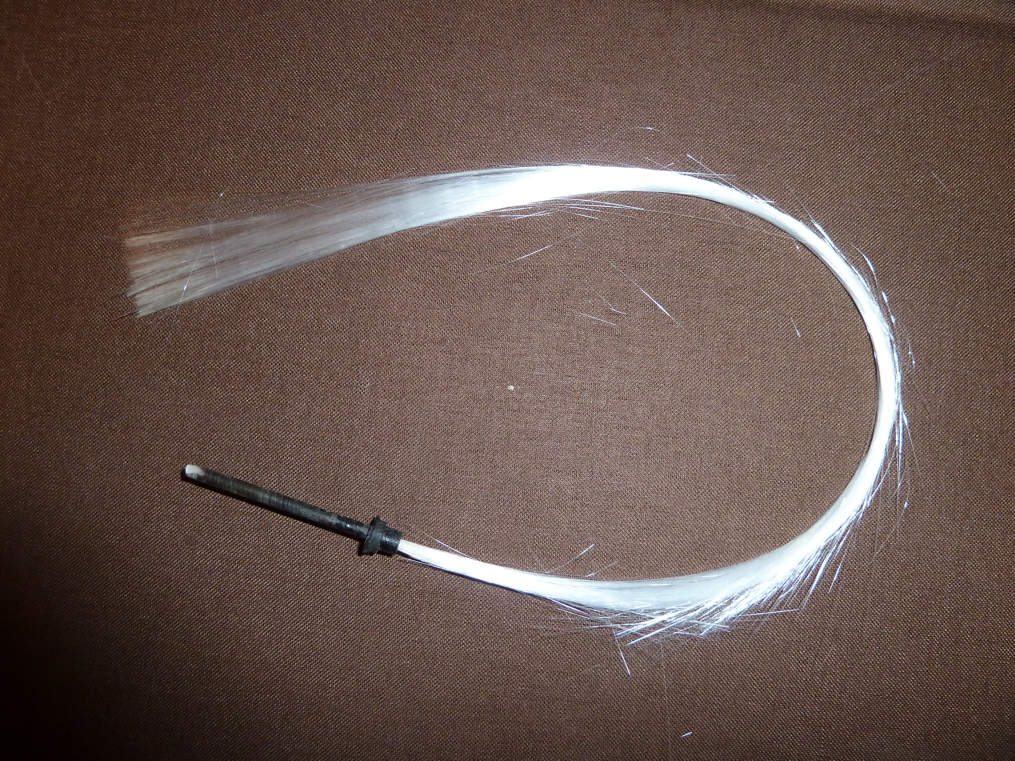
 The first concept of using coherent fibre-optical bundles to transmit images, ie. the ‘Fibrescope’ was devised by Harold H Hopkins and reported in 1954 1. Following this, fibreoptic endoscopes were in routine use in gastroenterology by the late 1970s. The image on the right is of a bundle of early glass fibre-optic lengths.
In 1964, Victor Marshall 2 of New York proposed the use of a flexible tube that could be manipulated within the body cavities to get into areas that were otherwise inaccessible. This 9Fr "ureteroscope" did not have any directional control but was passed into the kidney via an open ureterotomy in October 1960 and via a rigid cystoscope sheath up the ureter in May 1962.
Tsuchida and Sugawara in 1973 3 built the first flexible cystoscope that could visualise the inside of the bladder neck and provide a range of views, due to its ability to deflect at the tip. At 7mm diameter, it had a 90° deflection (each way), but this did not seem to convince urologists to explore further and development ceased.
In 1981 Harry Wilbur 4 used an 18FG choledochoscope with a 180° angle to inspect the lower urinary tract and discovered that it could potentially alter the dynamics of diagnostic urology. He was able to perform retrograde pyelography via a pre-existing suprapubic cystostomy, retrieve stents from ureters, and perform bladder biopsies and cauterisation. He emphasised using the surgeons’ existing choledochoscopes to limit expense in the urology department.
 Across in Germany the following year, 1982, Peter Burchardt used a 20FG flexible rubber choledochoscope with an arc view of 200° (100° deflection each way) to perform cystoscopy, urethroscopy or pyeloscopy; all with one instrument. He highlighted the ease with which flexible scopes can be used in bedridden patients in the supine position.
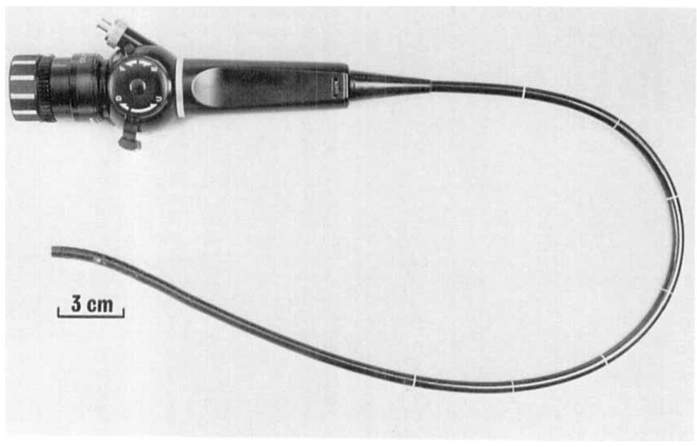
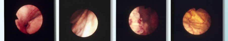
When approached, in 1980 by Stuart Greengrass of Olympus-KeyMed concerning the potential of fibre-optic cystoscopy, the BAUS Instruments Committee expressed little interest but, in August 1980, a prototype Olympus fibre-optic 16FG bronchoscope was evaluated for cystoscopy at Freeman Hospital, Newcastle upon Tyne. (image right is an Olympus BF fibre scope c.1983). The clarity of vision and ease of use through the urethra confirmed the potential. Photographs (below) of the prostatic urethra and bladder interior were taken through an Olympus 16FG bronchoscope at Freeman Hospital by Stuart Greengrass, in August 1980.
A prototype semi-flexible fibreoptic cystoscope was also developed, but was not successful due to its limited distal flexibility and poor insertion tube characteristics. A second prototype, the most sophisticated fibrescope insertion tube construction to date, was then designed to meet the particular requirements of bladder examination, namely, to visualise all aspects of the interior of a "domed’"sphere which required a much longer articulating bending section, rather than the interior of a tubular structure such as a bronchus, bile duct or bowel where a shorter bending section was required.
 With three regions of progressive flexibility to provide the necessary torque and retain urethral comfort, together with further total distal deflection of 330 degrees (210O up and 120O down) ,this permitted clear and easy inspection of all areas of the bladder, including the trigone and bladder neck, the latter by a J-manoeuvre of the fully flexed instrument.
Agreement was reached that this second prototype should be evaluated in routine clinical practice in the outpatient day-case setting by the Urology departments of the Freeman Hospital and the Royal London hospital. The findings of both studies (by Philip Powell and Christopher Fowler) were reported to the annual meeting of the British Association of Urological Surgeons in 1984 6,7.
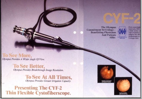
Further minor modifications to the design were incorporated to improve durability, handling and disinfection and the first successful purpose-designed flexible cystoscope, the Olympus CYF (right), was introduced into routine urology practice in the same year, thereby transforming diagnostic cystoscopy and the cystoscopic follow-up of people with superficial bladder tumours.
This article was written for the Museum of Urology by Mr Reg Hall, retired Consultant Urologist, Newcastle and Mr Stuart Greengrass former Director of R&D at Olympus-KeyMed - both pioneers of the flexible cystoscope
References
- Hopkins HH & Kapany NS. A Flexible Fibrescope, using Static Scanning. Nature. January 2, 1954; pp.39-41.
- Marshall VF. Fiber optics in urology. J Urol 1964; 91: 110.
- Tsuchida S & Sugawara H. A new flexible fibrecystoscope for visualization of the bladder neck. J Urol 1973;109(5): 830-831.
- Wilbur HJ. The flexible choledochoscope: a welcome addition to the urologic armametarium. J Urol 1981 126;(3): 380-381.
- Burchardt P. The flexible panendoscope. J Urol.1982; 127(3): 479–481
- Powell PH, V Manohar V, Ramsden PD, Hall RR. A flexible cystoscope. Br J Urol. 1984; 56(6)::622-4.
- Fowler CG, Badenoch DF, Thakar DR. Practical experience with flexible fibrescope cystoscopy in out-patients. Br J Urol 1984; 56(6): 618-21
← Back to Instruments & Equipment Room